I am back from de ICVSS2011 in Sicily (Italy), which was an amazing experience in several aspects attended by around of 160 Ph.D students and 15 speakers that work in the state-of-art in computer vision. Then, I want to share this experience in this post.
Before of ICVSS
The friday 8 of July Andrea Rueda and me traveled (in different flights) to Catania from Madrid. I arrived after of Andrea without know any of Italian but thanks of Google Maps and ask for the address showing a paper I finally find the accomodation place :)
The next day we went to Mount Etna which is imposing and impressive. At the end of this tour we can listen eruptions in the distance, the curious thing was that in the afternoon whereas waking in the center of Catania there was a volcanic ash rain clossing the airport all night. This was interesting but it was not a pleasant experience to feel the ash all over the body, because is like a very small and solid stone. Some of the students of ICVSS had to wait for the flight by the airport closed.
(volcanic ash cloud is comming)

(After of volcanic ash rain)

ICVSS starts
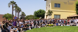
The sunday the bus was waitting for first group of ICVSS students that took us to the place of event. After of two hours of travel finally we are in Hotel Villagio Baia Samuele.
Poster sessions
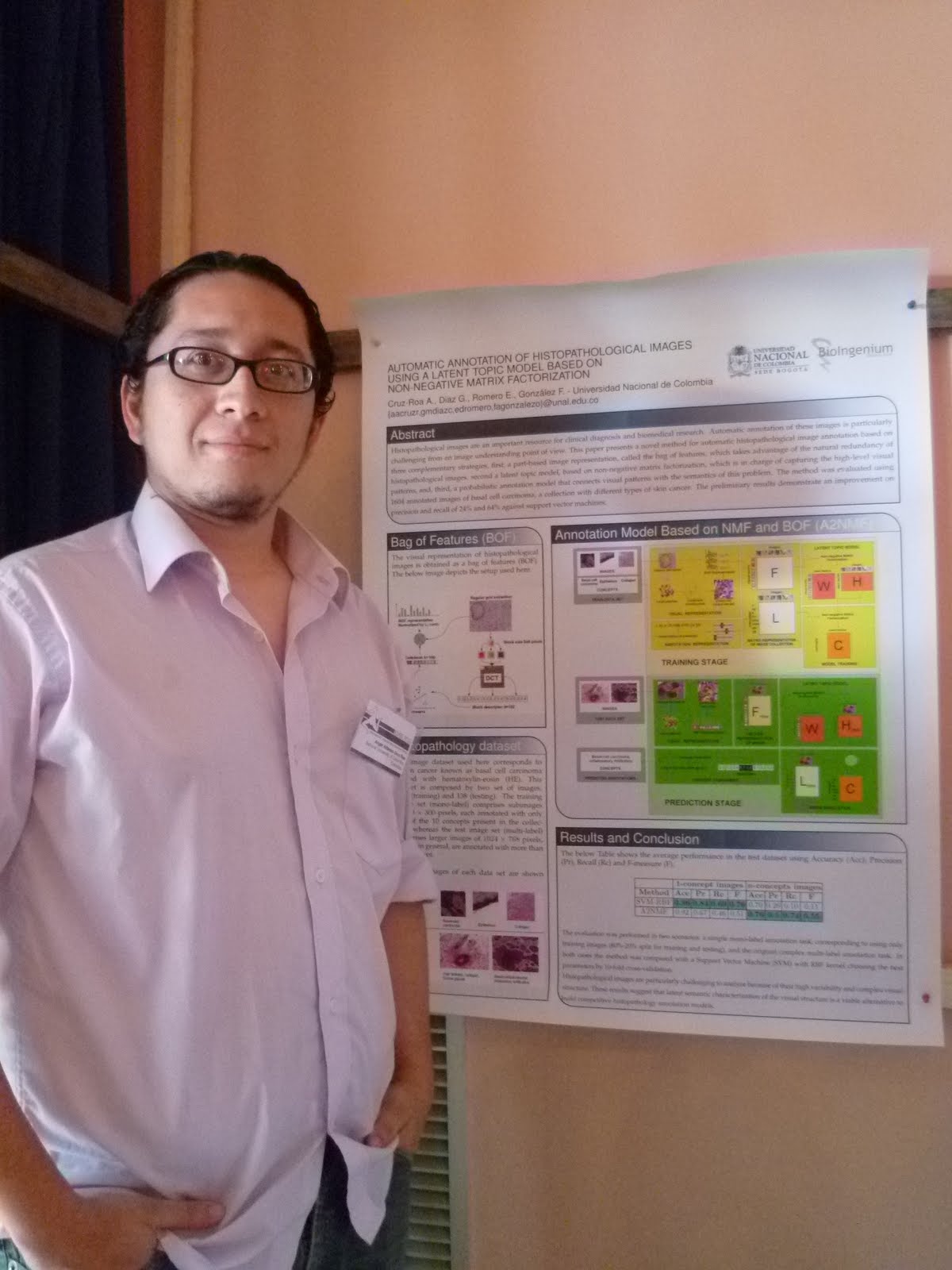
The Summer School starts the monday with the lectures all day with poster session of two hours at the final. The difficult degree was thanks of the hot inside of both floors where the poster session was performed. It was exhausting, I don't stop to talk in the all session without any drop of water :(
But were more the good things :D, many people like our proposed approach, in fact one of the things that the people likes was the way which we adapt Bag of Features approach (originally of Computer Vision area) in our context to represent Histopathological images.
One first conclusion is that I must improve the overall method diagram, still it is not clear enought. The description of Nonnegative Matrix Factorization problem must be included in the poster and the automatic annotation method also, step-by-step. Some persons also like the combination of BOF with NMF and the analysis that allows NMF for latent factors as a part-based representation, however few people didn´t know NMF and the people that know it said that is interesting because NMF has been applied in other areas for text and object recognition but is not common in biomedical images.
(The poster and me)
The common remarks about this work were:
It is a little weirdo the performance results obtained by SVM in accuracy and precision, in contrast with recall measure
- Maybe clarify that the classifiers are binaries and put how the performance measures were calculated over the average by image. Maybe use another measures like area under ROC curve, because the SVM model performance could be change according to threshold defined in the predicted labels.
- That is true, NMF doesn´t provide an unique solution. However the different solution are very closed. Maybe we must check this aspect, but in average NMF have similar results. This could be included in the way to present the results in future experiments. Nonnegative Tensor Factorization is a generalization of NMF that guarantees an unique solution, we must review and test this method.
- A: This database is not publicly yet, we are working in release but this take some time. We have another database that is publicly available of histology with images of tissues annotated by four concepts (http://www.informed.unal.edu.co/histologyDS/).
Are you going to test their method with other (similar and publicly) datasets?
- Yes, we must test with another datasets in histopathology images with several annotation by image. In fact also we can try with other kind of biomedical images, e.g. radiological images like ImageClef Medical database.
How the dataset was built? Who performs the annotations?
- A set of samples of basal cell carcinoma stained with Hematoxylin-eosin were digitalized from microscopy at the same magnification, which were globally annotated by an expert physician in pathology.
- Because NMF allows provide an interpretability layer of intermediate representation of images in latent space as a part-based representation in additive terms and not in terms of sums and substractions. The image, the concept or the latent factor is represented with mixtures in additive way of parts.
End of this part...
Interesting!! Nice post :)
ReplyDelete